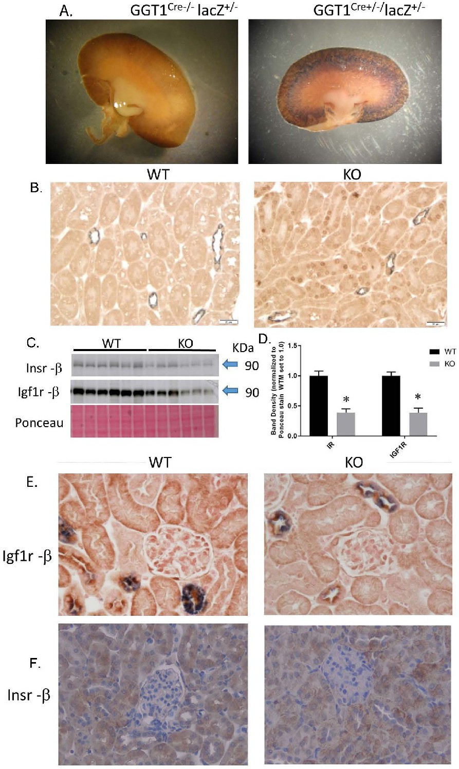
Fig. 1. Characterization of Insr/Igf1r PT-targeted dual KO. A. β -galactosidase-generated staining in a Cre-negative (left) and a Cre-positive (right) kidney cross-sections obtained from a mating between transgenic GGT1Cre+/- mice and lacZ reporter mice. B. Representative immunohistochemistry (IHC, brown stain) for nuclear Cre-recombinase in cortex sections from WT (left) and KO (right) mice; (gray stain; aquaporin-2 as a marker for collecting duct/connecting tubule). C. Western blotting for insulin receptor (β-subunit) and IGF1 receptor (β-subunit) in whole-kidney homogenates from WT and KO mice (n = 6/genotype); D. band densities for receptor blots normalized to Ponceau staining; E. IHC for Igf1r (β-subunit, brown) in the cortex of WT (left) and KO (right) mice; gray staining is aquaporin-2; F. IHC for Insr (β-subunit) in WT (left) and KO (right) mice; *indicates a significant (p<0.05) difference between WT and KO by unpaired t-test.